|
How to meet the right partner during brain wiring
Agi, Kulkarni et al., 2020, Curr. Op. Neurobiol.
Cover Caption :
Peters Rule!
|
|
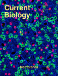
Rab GTPases and Membrane Trafficking in Neurodegeneration
Kiral et al., 2018, Curr. Biol.
Cover Caption :
In the cover image of the developing fly eye, extensive subcellular compartmentalization is revealed via antibody-mediated detection of membrane-bound organelles; the endoplasmic reticulum is shown in blue, the endolysosomal system in green, and nuclei in magenta. Image courtesy of Friederike Kohrs, Jen Jin, and Ridvan Kiral.
|
|
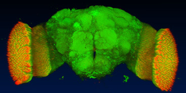
The synaptic vesicle SNARE neuronal Synaptobrevin promotes endolysosomal degradation and prevents neurodegeneration
Haberman et al., 2012, Journal of Cell Biology 196, 261-276
Cover Caption :
A 3D visualization shows the photoreceptor projections in the adult Drosophila brain. Haberman et al. reveal that, in addition to regulating synaptic vesicle exocytosis, the SNARE protein Synaptobrevin also promotes endolysosomal degradation. Loss of this degradation mechanism in photoreceptors leads to adult-onset degeneration.
|
|
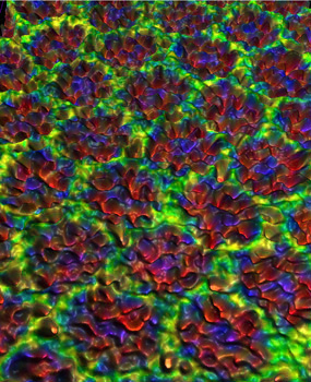
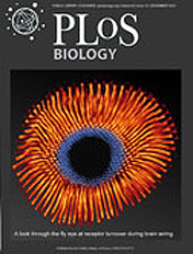
Guidance Receptor Degradation is Required for Neuronal Connectivity in the Drosophila Nervous System
Williamson et al., 2010, PLoS Biology 8(12): e1000553.
Cover Caption :
The picture shows a preparation of the eye of the fruit fly, Drosophila melanogaster. The eye is imaged from the inside and fluorescently labeled for the light sensitive rhabdomeres using Phalloidin. A confocal maximum projection visualization was false-colored to distinguish rhabdomere cross-sections (center, blue) from tangential sections (orange). The Drosophila eye forms a stereotyped pattern during development and extends axons into the brain using specialized guidance cues. In this issue of PLoS Biology, Williamson et al. propose that a neuron-specific protein degradation pathway is required for the spatiotemoral regulation of guidance receptors during development of the Drosophila visual system.
|
|
A dual function of V0-ATPase a1 provides an endolysosomal degradation mechanism in Drosophila photoreceptors
Williamson et al., 2010, J. Cell. Biol. 189, 885-99.
Cover Caption :
In the Drosophila visual system, photoreceptor neurons (green) project toward the optic lobe and form synaptic connections in the neuropil (red). Nuclei are labeled blue. Williamson et al. show that the neuron-specific v-ATPase subunit v100 has a dual function in sorting and degrading proteins through the endolysosomale pathway, protecting the photoreceptors from neurodegeneration.
|
|
The v-ATPase V0 subunit a1 is required for a late step in
synaptic vesicle fusion
Hiesinger et al., 2005, Cell 121, 607-620.
Cover Caption :
The cover shows a confocal microscopy heightfield visualization of synapses in the first optic neuropil of the adult fly, labeled with the synaptic markers Vha100-1 (red), nc82 (green), and DPAK (blue). Vha100-1 is a v-ATPase V0 complex component found at synapses of the CNS as well as neuromuscular junctions. A possible role of the V0 complex in membrane fusion independent of v-ATPase function in neurons is a long-standing controversy. Hiesinger et al. report on pp. (607-620) a synaptic requirement of Vha100-1 for a late step in exocytosis (pp. 303-313).
|
|
Endophilin acts after
Synaptic Vesicle Fission in Drosophila Photoreceptor Terminals.
Fabian-Fine et al., 2003, J. Neurosci 23, 10732-44
Cover Caption : Three-dimensional volume rendering of the first
optic neuropil, or lamina, of the Drosophila optic lobe labeled with
anti-endophilin (Endo; green) and anti-synaptobrevin (Syb; red). Precise
modules of this neuropil, called cartridges, comprising six photoreceptor
terminals that innervate lamina monopolar cell interneurons, are
immunoreactive for Endo and Syb (orange/yellow). Note, however, that Endo is
also present in unidentified cells of the lamina cortex surmounting the lamina.
(This image was prepared by P. Robin Hiesinger.) For details, see the article
by Fabian-Fine et al. in this issue (pages 10732-10474).
|
|
Drosophila VAP-33A directs bouton formation and neuromuscular junctions in a dosage-dependent manner.
Pennetta et al., 2002, Neuron 35, 291-306.
Cover Caption : The cover shows the end of a neuronal branch
(foreground) at the Drosophila neuromuscular junction (background). Boutons
are stained with an antibody against DVAP-33A (yellow to brown), which is
excluded from active zones (purple). DVAP-33A is enriched at the neck of
budding boutons. The visualization of the surface and underlying
triangularized wireframe (top left) are merged with a qualitative volume
rendering of a bouton from which the top is removed. Functional
characterization of DVAP-33A using these visualization techniques shows that
DVAP-33A is a microtubule-interacting protein and tightly regulates bouton
budding at neuromuscular junctions in a dosage-dependent manner. For further
details, see the article by Pennetta et al. (pp. 291-306 in this issue).
|
|
Neuropil pattern
formation and regulation of cell adhesion molecules during Drosophila
optic lobe development depend on synaptobrevin.
Hiesinger et al., 1999, J. Neurosci. 19, 7548-7556
Cover Caption : Three-dimensional reconstruction of a midpupal
Drosophila optic lobe. The green channel shows inactive tetanus toxin light
chain expression driven by the enhancer Gal4 line Mz1369. A staining of
neuropil structures with an antibody against the cell adhesion molecule
IrreC-rst is shown in red. Expression of active tetanus toxin light chain
under control of Mz1369 results in a disturbance of neuropil fine structure
as well as an upregulation of IrreC-rst immunoreactivity. For details, see
the article by Hiesinger et al., in this issue (pages 7548-7556).
|
|
Shar-pei
mediates cell proliferation arrest during tissue growth in Drosophila.
Kango-Singh et al., 2002, Development 129(24), 5719-30. (cover
picture by Georg Halder)
Cover Caption : Pupal retina of Drosophila melanogaster imaged by
confocal microscopy. Cell outlines of cone and pigment cells are visualized
by Discs-large expression (green) and a deeper optical section shows
photoreceptor nuclei labeled with TOPRO (blue). Circular structures are
ommatidial cell clusters composed of four cone cells and eight photoreceptor
cells that are surrounded by pigment cells. The retina is mosaic for shar-pei
mutant and wild-type cell clones. Wild-type cells express GFP (red), while
mutant cells do not. shar-pei mutant cells show excessive cell proliferation.
For further details see article by M. Kango-Singh, R. Nolo, C. Tao, P.
Verstreken, P. R. Hiesinger, H. J. Bellen and G. Halder in this issue, pp.
5719-5730.
|
|
|
Some German Blurb on
3D Visualization...
...a bit in the style of "yes, flies do have a brain and computers are fun, kids."
You may not want to read this...
Hiesinger and Fischbach (2000). BioSpektrum. 5/2000, 408-412.
|